
Almost every week, a patient over fifty mentions in passing that their mother had “that thing where she couldn’t read anymore.” That thing is usually age-related macular degeneration, and the next ten minutes of the appointment matter more than most people realize.
AMD is the leading cause of central vision loss in adults over fifty in the United States. It is partly genetic, partly lifestyle, and almost entirely silent in its earliest, most treatable stages. The patients we worry about most are the ones who say, “Well, I see fine.” So do most early AMD patients — for years.
What “family history” actually means
This is the conversation we want to have with you. Having a parent or sibling with AMD meaningfully raises your own risk — studies generally put it at roughly two to four times the baseline. Certain inherited gene variants are among the strongest known predictors of who progresses to advanced disease. But genetics is not destiny, and much of what determines your outcome comes down to factors you can actually change — and to whether anyone is watching your retinas closely enough to catch the earliest changes.
So a useful first question at your exam is not “do I have it?” but rather: given what you know about my family and my own retinas today, what is my risk over the next ten years, and what can I do about it?
Why early detection is the entire game
AMD has two main forms. Dry AMD is the slow form — the macula thins, small yellow deposits called drusen accumulate under the retina, and central vision dims gradually. The large majority of AMD is dry. Wet AMD is the dangerous form — abnormal blood vessels grow under the retina and leak, and central vision can drop in days to weeks. Wet AMD is treatable with injected medications (anti-VEGF drugs, given by a retina specialist), and outcomes are dramatically better when treatment starts before significant scarring forms.
The math, then: catching AMD in the dry phase, and catching the conversion to wet the moment it happens, is the single most consequential thing your eye doctor can do for you. Annual exams with retinal imaging make that possible.
The imaging that catches it early — Optomap and OCT
Two technologies in our office do most of the heavy lifting here, and they complement each other.
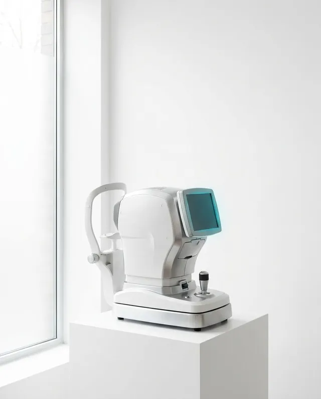
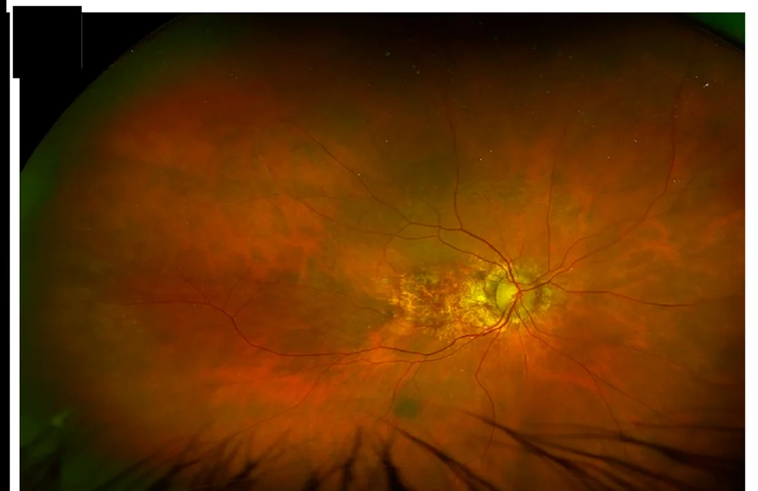
Optomap® ultra-widefield retinal imaging captures roughly 200 degrees of the retina in a single, dilation-free scan. We document the macula in a high-resolution image and compare it side by side with prior years. Early drusen, pigment changes, and the first hints of atrophy show up in these images long before they affect reading vision. The comparison over time is the point — a single photo tells us where you are; a series tells us where you are headed.
Optical coherence tomography (OCT) is the other half of the picture. Where Optomap gives us a photograph of the retina’s surface, OCT takes a cross-section through it — showing each microscopic layer of the macula in fine detail. It is the test that catches the conversion from dry to wet AMD, often before a patient notices anything at all, because a small pocket of fluid under the retina is visible on OCT before it distorts vision. For patients with a family history of AMD, this scan earns its place in the exam.
Both are quick, painless, and done right here in Waynesville. If treatment is ever needed, we coordinate the referral to a retina specialist and stay involved in your care.
The Amsler grid — at home, weekly
If you have any AMD risk, this is a small habit worth building. The Amsler grid is a free, paper-based home check that helps catch the conversion from dry to wet AMD between visits. Look at the center dot, one eye at a time, holding the grid at normal reading distance with your reading glasses on if you use them. Any lines that look wavy, broken, or that have blank spots — that is the day to call us, not next month. Tell us you have new distortion on your Amsler grid; that phrase gets you in fast.
What you can actually do
This is the hopeful part. The AMD risk factors you can modify are among the best-studied in eye care.
- Don’t smoke. Smoking is the strongest modifiable risk factor and substantially accelerates AMD. If you smoke, quitting matters more for your eyes than any vitamin you can buy.
- AREDS2 vitamins — if your retinas show the right findings. The large AREDS and AREDS2 trials showed that a specific formula of lutein, zeaxanthin, vitamins C and E, zinc, and copper slows the progression of intermediate AMD to advanced disease by roughly a quarter. It is not a preventive for healthy eyes — it is a slowing agent for the right patient. Whether it applies to you depends on what your imaging shows, which is exactly the conversation to have at your exam.
- Eat dark leafy greens. Spinach, kale, collards — high in lutein and zeaxanthin, the pigments that concentrate in the macula. Twice a week, minimum.
- Wear sunglasses outside. Long-term sun exposure adds to wear and tear on the retina, and mountain sun at elevation is not gentle. Proper UV-blocking sunglasses are cheap insurance, and our optical carries plenty of options — including prescription and polarized.
- Mind your cardiovascular health. Anything good for your heart is good for the small vessels feeding your retina: blood pressure, cholesterol, blood sugar.
- Keep the annual exam. It is the cheapest, most effective way to catch problems early. Put it on the calendar every year.
The questions to ask at your next exam
Bring this list. We mean it — patients who ask direct questions get better care everywhere, not just here.
- What do my retinal photographs and OCT scan show today?
- Is there any early drusen visible? If so, where on the macula?
- Should I have a baseline Optomap and OCT on file for comparison going forward?
- Given my family history, what is my risk over the next ten years?
- Should I be taking an AREDS2 formula?
- How often should I check the Amsler grid at home?
Several of our doctors follow AMD patients closely — Dr. Krempecki in particular has a clinical focus on macular degeneration, along with diabetic eye disease and glaucoma — so if AMD runs in your family, you will be in experienced hands whichever doctor you see.
When to call us sooner
New distortion is the symptom that outranks the calendar. If straight lines start to bend, a smudge or gray spot appears in your central vision, or one eye’s vision drops noticeably over days — call (828) 456-8361 that day and tell us what you are seeing. Fast action is what preserves vision in wet AMD.
Booking an exam
If you have a parent or sibling with AMD and have not had a comprehensive exam in the last twelve months, this is exactly what we want to see you for. Schedule online anytime, or call (828) 456-8361. Mention the family history when you book so we plan the right imaging into your visit.